Electrospun Scaffolds for ORL Reconstruction
The successful outcome of reconstructive procedures for the ear, nose and throat (ORL) is defined not only by the functional properties of the reconstructed area, but also by aesthetic aspects. Especially in the case of trauma or large scale tumour resections, sufficient autologous cartilage material for functional and aesthetic reconstruction is often lacking. Due to the limited regenerative capacity of cartilage itself, there is a strong demand for novel therapeutic options for these patients.
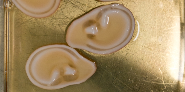
Therefore, this research aims to develop a novel method for producing patient-specific cartilage scaffolds for ORL reconstruction surgeries, using a method called electrospinning. It can be described as forming fibres by preferential deposition of a solidifying liquid or gel on a surface through the application of a very high electrical potential (20-30kV) between the spinning outlet nozzle and the conductive receiving surface.
This receiving surface usually consists of either a flat collector plate, or a rotating mandrel, which is why conventional electrospinning forms sheet-like or tubular structures given by the collector geometry. With fast collector movement, a certain degree of fibre alignment can be achieved which leads to improved tensile strength of the construct in fibre direction.
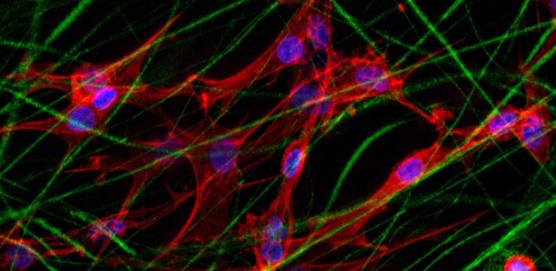
However these conventional methods are not suitable for the described project aims. Therefore, new strategies are being developed for generating more complex 3-dimensional shapes suitable for ORL reconstruction purposes. Further emphasis is put on fine-tuning the scaffold microstructure, in order to not only improve mechanical stability, but also to optimize adhesion and metabolism of seeded chondrocytes, hence allowing to create a living tissue substitute.
Contact
Head of Institute for Biomechanics
Institut für Biomechanik
Gloriastrasse 37/ 39
8092
Zürich
Switzerland